What is canine cruciate disease?
Cranial cruciate ligament disease (CCLD) is the leading cause of hindlimb lameness in the dog (Powers et al, 2005). The cranial cruciate ligament (CCL) is the canine-equivalent of the human anterior cruciate ligament (ACL). The CCL is the primary passive stabiliser of the canine stifle joint, it’s craniomedial band taut during flexion, while the caudolateral band and lateral collateral ligament relax to allow for tibial internal rotation (de Rooster et al, 2006). In addition to stifle joint stability, these ligaments provide mechanoreceptor and proprioceptive input (de Rooster et al, 2006).
Injuries of the human ACL typically result from acute, non-contact trauma involving deceleration/acceleration in combination with a knee valgus load (Logerstedt et al, 2010). Acute CCL rupture in dogs can result from a single high-load traumatic event, however, in most cases, CCLD is the result of normal forces placed on a ligament that has undergone chronic degenerative change (Jerram and Walker, 2003; Hayashi et al, 2003). This process is progressive, involving gradual degeneration of the CCL, stifle joint inflammation, and leads to partial and eventually, complete ligament rupture (Hayashi et al, 2003). Secondary osteoarthritis frequently develops (de Bruin et al, 2007).
Aetiopathogenesis
Aetiopathogenesis2003). This process is progressive, involving gradual degeneration of the CCL, stifle joint inflammation, and leads to partial and eventually, complete ligament rupture (Hayashi et al, 2003). Secondary osteoarthritis frequently develops (de Bruin et al, 2007).
A number of risk factors for CCLD have been identified, including:
An increased prevalence for CCLD in female dogs, and in neutered over sexually-intact dogs (Slauterbeck et al, 2004).
Obesity increases the risk for CCLD, with overweight dogs demonstrating altered kinematics relative to healthy-weight dogs (Lapman et al, 2003; Bray et al, 2013).
Medial patella luxation, genu varum, and conformational variations such as a straight stifle joint angle, narrow distal femoral intercondylar notch, and steep tibial plateau angle (TPA) have all been associated with CCLD (Haynes et al, 2015; Schwandt et al, 2006, Mostafa et al, 2009).
Material properties of the CCL decrease with age, increasing the risk of rupture in dogs >; 4 years, especially in ‘high risk’ breeds (Witsberger et al, 2008; Duval et al, 1999).
Breed variation in biomechanical properties of the CCL, with significantly lower load-to-failure properties reported in some breeds (Wingfield et al, 2000a; Wingfield et al, 2000b).
Immune-mediated conditions of the stifle (Doom et al, 2008).
Of note, despite the multiple risk factors for CCLD, it remains unclear which of these lead to disease initiation (Hayashi, 2018).
Surgical techniques for Cranial Cruciate Ligament repair
Lateral Fabellar Stabilisation (LFS)
LFS (also known as De Angelis procedure) is an extracapsular repair technique (De Angelis and Lau, 1970). The faballae are small sesamoid bones, one for each tendon of the two heads of gastrocnemius (Kowaleski et al, 2018). With LFS, the aim is to passively stabilise the stifle and prevent cranial (anterior) tibial translation with respect to the femur, of which would normally be prevented by the intact cranial cruciate ligament (Heffron and Campbell, 1978). A nonabsorbable synthetic suture material is passed around the lateral fabella and back through one or two bone tunnels made in the tibial tuberosity (Casale and McCarthy, 2009). The suture is held taut by a knot or crimp clamp (Lotsikas et al, 2013). The suture material eventually stretches or breaks, but the temporary stabilisation it provides allows for the formation of organised scar tissue, ultimately leading to long-term stability (Warzee et al, 2001).
The advantages of LFS are its’ relative technical ease and shorter surgical time, however, if there is low tensile strength or increased creep of the suture material, this can lead to insufficient or excessive scar tissue formation and joint laxity (Christopher et al, 2013).
Corrective Osteotomies
The Tibial Plateau Leveling Osteotomy (TPLO) and Tibial Tuberosity Advancement (TTA) are the two most common corrective osteotomy techniques performed (Conzemius et al, 2005).
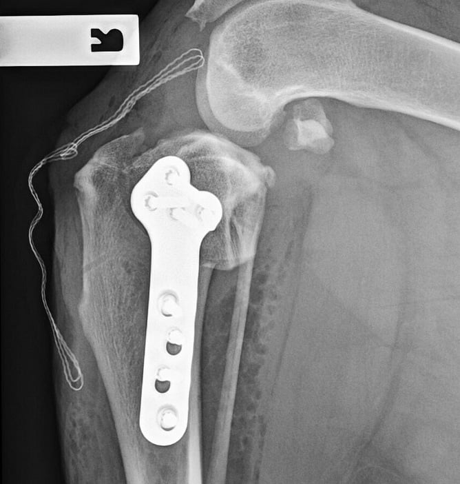
The TPLO involves cutting a curved osteotomy through the proximal tibia and rotating the tibial plateau segment to achieve a lower TPA (Lotsikas et al, 2013). After rotation, a plate and screws are applied to stabilise the osteotomised segment (Kowaleski et al, 2018). The aim is to neutralise cranial drawer by leveling the tibial surface so it is perpendicular to the load applied during weightbearing (Kowaleski et al, 2018). The TPLO alters biomechanics by placing additional reliance on the caudal cruciate ligament and muscular stabilisers of the stifle.
The TTA involves an osteotomy caudal to the tibial tuberosity and advancing of the tibial tuberosity so the patellar tendon is perpendicular to the tibial plateau. A titanium cage and plate hold the advanced portion of bone in place (Kowaleski et al, 2012). The TTA neutralises cranial drawer by altering forces through the stifle to be parallel to the patellar tendon, providing a biomechanical advantage to the quadriceps muscles (Guerrero et al, 2011). This procedure is less technically demanding, but has a higher incidence of post- operative patella tendonitis and meniscal tears compared with TPLO (Hurt et al, 2011).
More recent studies have shown that TPLO and TTA are superior to extracapsular stabilisation techniques, with TPLO providing better long-term radiographic and functional outcomes than TTA (Krotscheck et al, 2016; Moore et al, 2020).
Evidence for Physiotherapy in Canine Cruciate Disease
Physiotherapy following CCL stabilisation has been reported in three studies (Marsolais et al, 2002; Monk et al, 2006; Jerre, 2009). Marsolais and colleagues (2002) assessed the effectiveness of passive range of motion (PROM), massage, walking and swimming following extracapsular stifle stabilisation and found improvements in peak vertical force and vertical impulse six months after surgery. Monk et al (2006) compared the physiotherapy modalities of cold, massage, PROM, underwater treadmill, and progressive functional exercises to a home-exercise programme in dogs following TPLO. At six weeks thigh circumference and stifle PROM was significantly greater in the physiotherapy group compared with controls (Monk et al, 2006).
Jerre (2009) compared two groups, in which the control group received a graduated walking programme and instructions for massage and stretching were provided to the owner. In addition to this, the treatment group received swimming and transcutaneous electrical nerve stimulation. No significantly detectable differences were reported between the two groups.
It has been suggested that physiotherapy following CCL stabilisation may be more important in terms of patient overall outcome than choice of surgical technique (Marcellin-Little and Arnoldy, 2018).